A study on stress responses by co-creation of liquid-liquid phase separation and selective autophagy
Department of Organ and Cell Physiology, Juntendo University Graduate School of Medicine Dr. Masaaki Komatsu

A study on stress responses by co-creation of liquid-liquid phase separation and selective autophagy
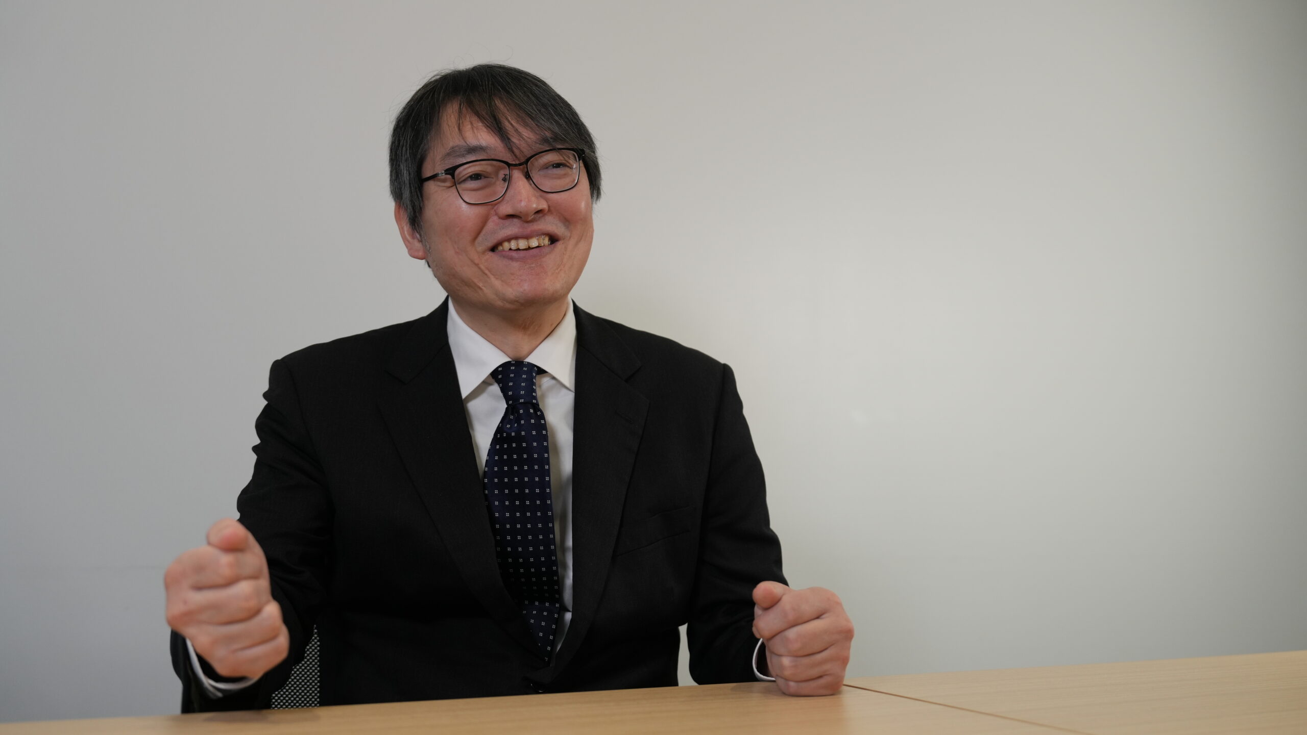
Selective autophagy, a process that targets and degrades denatured proteins and dysfunctional organelles, has become a major focus of research worldwide. Professor Masaaki Komatsu, from the Department of Organ and Cell Physiology, made history by creating the world’s first conditional autophagy-deficient mice, establishing himself as a leading figure in the field. His latest research explores liquid-liquid phase separation. We spoke with Professor Komatsu for an interview to learn more about the current state of autophagy research and its future directions.
Autophagy is a cellular process that degrades unnecessary proteins and damaged organelles, repurposing their components for energy production and macromolecule synthesis. Autophagy gained widespread recognition in 2016 when Dr. Yoshinori Ohsumi of the Tokyo Institute of Technology (now Institute of Science Tokyo) was awarded the Nobel Prize in Physiology or Medicine for elucidating its mechanism.
The human body is composed of approximately 38 trillion cells, each equipped with an autophagy mechanism. Autophagy is a ‘self-eating’ process that helps maintain intracellular proteostasis and organellostasis by routinely removing unnecessary components. This process involves the formation of a membrane that engulfs parts of the cell, including organelles, to form a structure called an autophagosome. The autophagosome then fuses with lysosomes, which degrade the engulfed cellular components.
Research on autophagy dates back to the 1960s when Dr. Christian de Duve, a Belgian biochemist, identified autophagy as a self-degradative mechanism. However, fully elucidating its mechanism required identifying the genes involved, leading to more than 30 years of morphological studies, including electron microscopy observations as well as biochemical analyses using rat livers and primary cultured hepatocytes.
The next breakthrough came with the work of Professor Yoshinori Ohsumi. He demonstrated autophagy in budding yeast, a species commonly used in bread making, and in 1993 successfully identified a series of ATG genes essential for autophagy. His research led to the discovery of more than a dozen genes crucial for autophagosome membrane formation, significantly advancing the field.
In the 2000s, autophagy-deficient knockout mice were developed, leading to significant insights into the fundamental functions of autophagy. It became evident that autophagy serves as an ‘ultimate survival strategy’ by degrading cellular proteins to generate amino acids for energy during periods of nutritional deprivation, such as fasting. Additionally, research demonstrated that autophagy operates continuously at low levels even under nutrient-rich conditions, playing a crucial role in maintaining intracellular metabolic homeostasis.
In the 2010s, studies revealed that autophagy deficiency results in the accumulation of misfolded proteins and dysfunctional organelles, contributing to neurodegenerative diseases and cancer. Parkinson’s disease, in particular, was found to have a strong association with autophagy. The causative genes PINK1 and Parkin regulate the autophagic degradation of damaged mitochondria, thereby maintaining mitochondrial quality. However, when this function is impaired, defective mitochondria accumulate, contributing to the progression of Parkinson’s disease.
Recent advances in research have shown that autophagy exhibits ‘selectivity,’ enabling the targeted degradation of specific misfolded proteins and dysfunctional organelles. This suggests that the accumulation of substrates that should have been selectively degraded by autophagy may contribute to disease development. Beyond its fundamental role in cell survival and homeostasis, autophagy is also essential for stem cell maintenance, aging regulation, and adaptation to environmental stress. A deeper understanding of these functions requires elucidating the mechanisms underlying selective autophagy.
To elucidate the function of selective autophagy, our research team has been conducting pathophysiological analyses using genetically modified mice for the past 25 years. In 2005, we became the first in the world to successfully generate ‘conditional autophagy-deficient mice,’ which allow the selective deletion of the autophagy-related gene Atg7 in specific organs. Previously, systemic deletion of Atg genes resulted in embryonic lethality or death within one day after birth, making detailed analysis in specific organs, tissues, or adult mice impossible. By utilizing organ-specific knockout mice, we have been able to investigate the relationship between autophagy and various organs, such as the brain and liver. Our findings have demonstrated that autophagy inhibition is a key factor in the development of multiple diseases.
A crucial component in understanding selective autophagy is the intracellular protein p62. Misfolded proteins and dysfunctional organelles that require autophagic degradation are tagged with ubiquitin. Our research focuses on the relationship between ubiquitinated protein aggregates and p62.
In our 2005 study, we confirmed that ubiquitinated protein aggregates accumulate in Atg7-deficient mouse hepatocytes, leading to liver damage. The following year, we demonstrated that the absence of autophagy in neuronal cells resulted in neurodegenerative disease in mice. Then, in 2007, we revealed that p62 plays a critical role in the formation of ubiquitinated protein aggregates in the hepatocytes and neuronal cells of Atg7-deficient mice.

Inside cells, certain proteins and biomolecules spontaneously form liquid-like droplets through a process called liquid-liquid phase separation (LLPS). These droplets remain dynamic, allowing molecules to cluster while maintaining fluidity, enabling biochemical reactions and cellular regulation. LLPS helps organize cellular components without membranes, facilitating stress responses, RNA processing, and protein degradation. However, abnormal droplet formation or persistence can lead to neurodegenerative diseases and cancer.
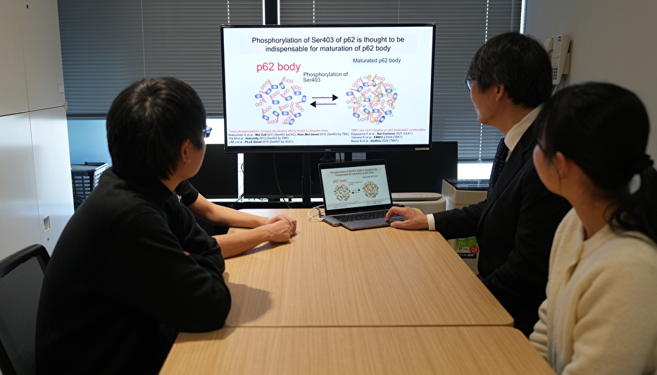
We discovered that under various stress conditions, the accumulation of misfolded and denatured proteins triggers their ubiquitination, which in turn leads to multivalent interactions with the intracellular protein p62, resulting in the formation of large droplet-like structures known as p62 bodies.
Furthermore, we discovered that p62 bodies are not just passive droplets degraded by autophagy, but they also have critical cellular functions. One such function is the activation of the KEAP1-NRF2 pathway, a major stress response mechanism. In response to stress, p62 bodies sequester KEAP1, a protein that normally facilitates the degradation of NRF2, a key transcription factor for oxidative stress responses. As a result, NRF2 is stabilized and activated. In other words, in response to cellular stress, accumulated ubiquitinated proteins are converted into p62 bodies, which in turn activate NRF2 to mitigate stress while simultaneously being degraded by autophagy to eliminate abnormal proteins. This reveals a rational mechanism that links stress adaptation with protein quality control.
The functional distinction between the stress-response role of p62 bodies and their degradation through autophagy highlights their context-dependent versatility. On one hand, the autophagic degradation of p62 condensates is crucial for maintaining proteostasis by clearing misfolded and ubiquitinated proteins. On the other hand, their participation in the KEAP1-NRF2 pathway demonstrates their regulatory role in oxidative stress adaptation. Despite this dual functionality, the mechanisms regulating the switch between these roles remain unclear. Furthermore, it has been revealed that the contents of p62 bodies vary depending on stress conditions, leading to changes in their function. Our research aims to understand the formation, composition, physical properties, dynamics, cellular functions, and autophagic degradation of p62 bodies in response to various stress conditions.
This research project also aims to establish an international research hub, leveraging Juntendo University’s expertise in autophagy to train young researchers and advance the field. Through collaborations with six world-leading autophagy research groups from Japan and abroad, two bioimaging teams, and three clinical groups specializing in blood cancer, neurodegenerative diseases, and metabolic and endocrine disorders, the initiative aims to how autophagy maintains intracellular homeostasis and the pathological processes underlying its dysfunction.
The research team includes scientists from Juntendo University, Hokkaido University, and Institute of Science Tokyo, as well as experts from China, the UK, and the US. By providing young researchers with opportunities to study abroad, the project aims to cultivate the next generation of leaders in autophagy research.”
Our primary goal is to steadily elucidate the functions of selective autophagy, a mission we are deeply committed to.
Specifically, we aim to comprehensively identify novel components of the p62 body in response to various stress conditions. To achieve this, we will employ an innovative purification method for the p62 body, along with a technique to inhibit selective autophagy. In collaboration with clinical researchers on our team, we will also focus on liver diseases and neurodegenerative diseases, where abnormalities in p62 body formation and degradation are believed to contribute to disease onset. Furthermore, we will investigate the dynamics of the p62 body using multiple mouse models of liver and neurodegenerative diseases, human three-dimensional liver organoids, and patient-derived specimens.
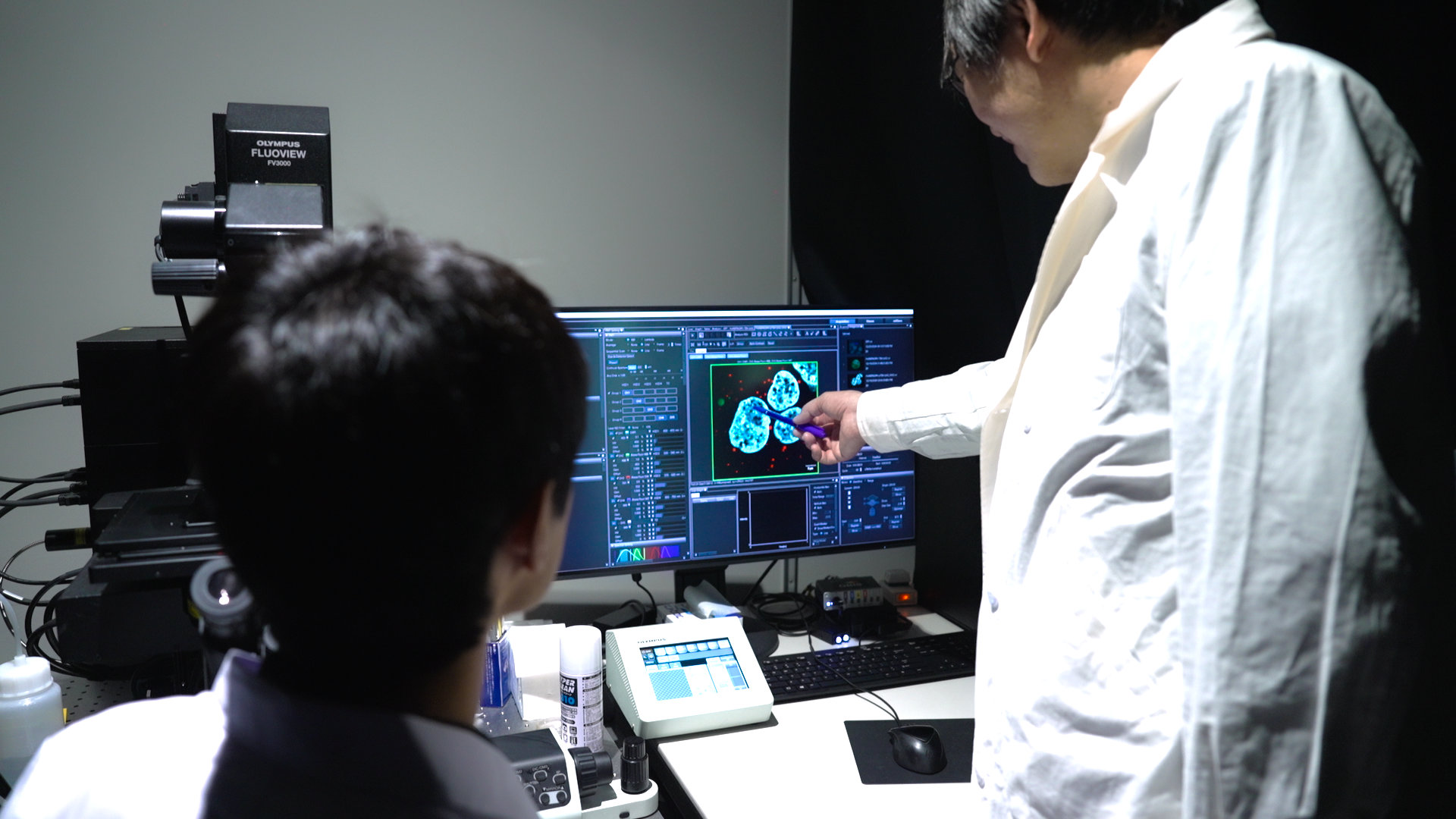
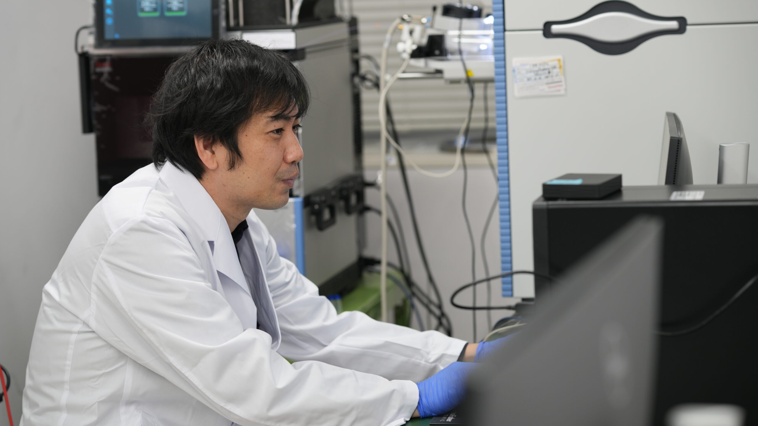
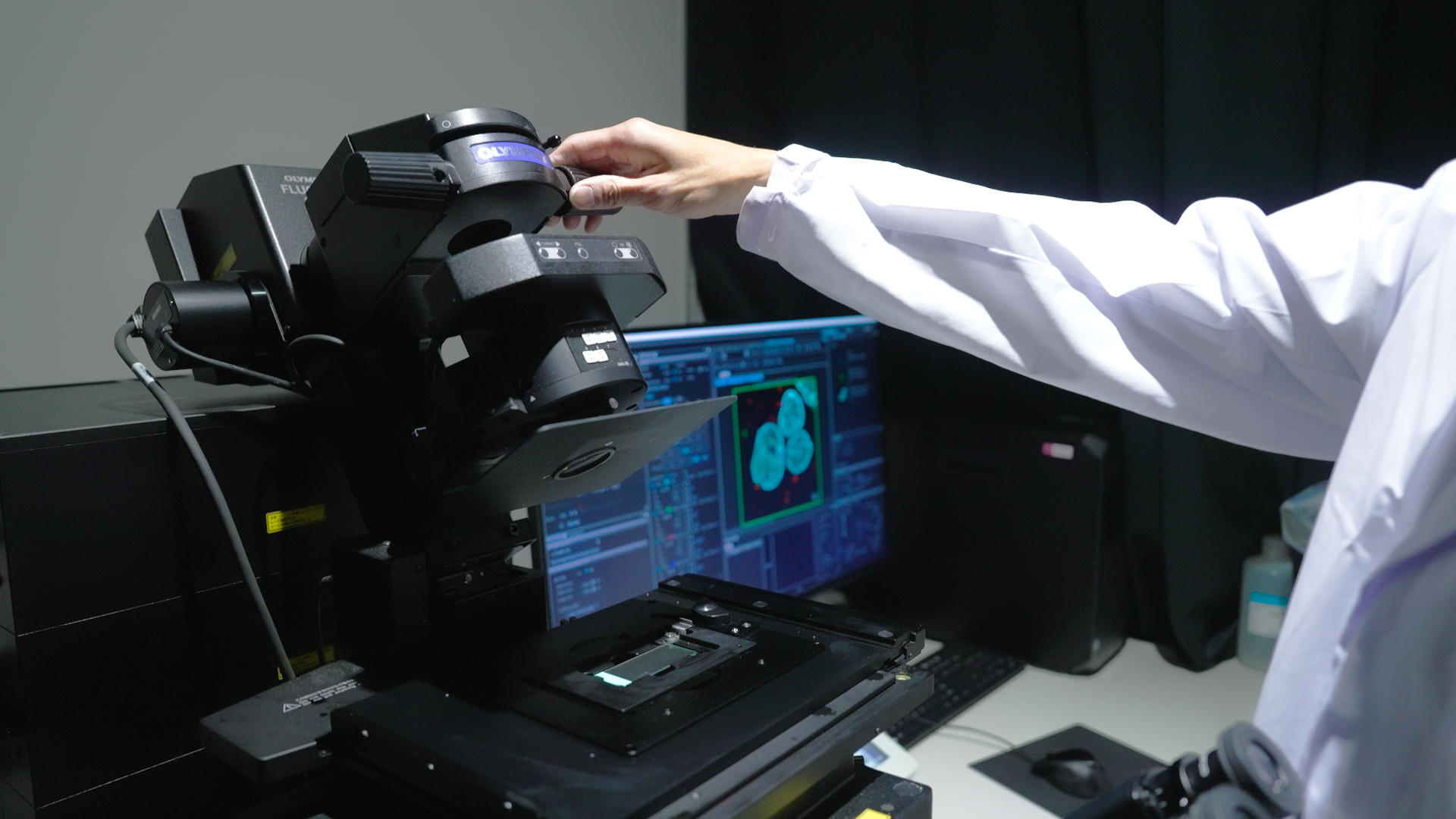
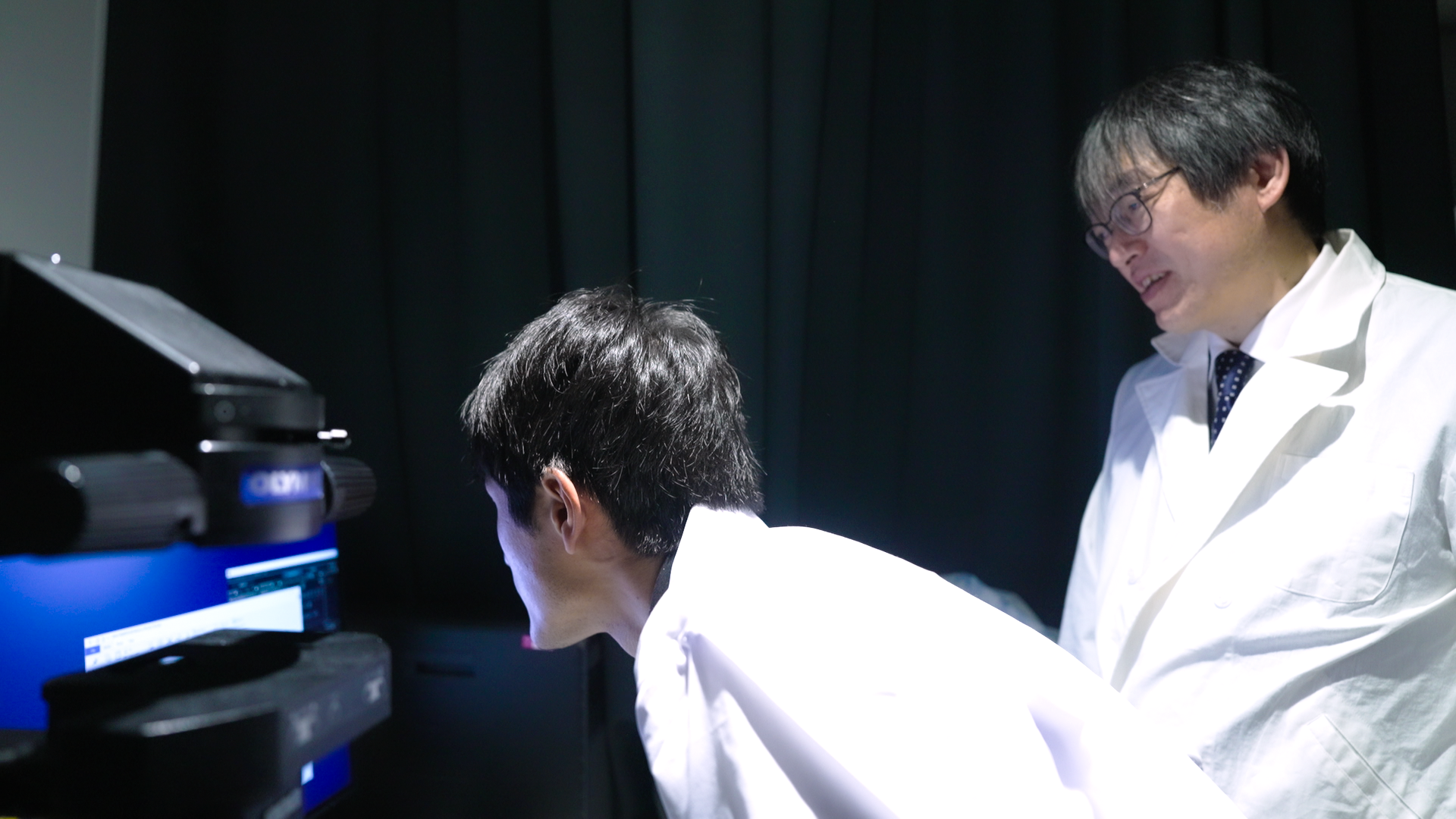
Juntendo University provides an optimal environment for this research. The university fosters a strong community of clinicians who actively support basic research and offers access to a large number of clinical cases of liver and neurodegenerative diseases, which align with our research focus. The university is home to the Autophagy Research Center, which is equipped with advanced mass spectrometers, as well as the Imaging Center, which provides access to state-of-the-art super-resolution and electron microscopes essential for morphological studies.
By leveraging the expertise of our team members and the cutting-edge resources available at our facilities, we strive to elucidate the function of selective autophagy and train the next generation of researchers in this field.
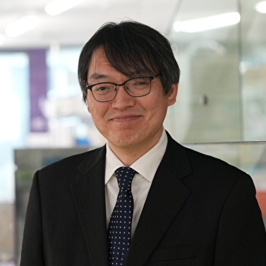
Professor
Department of Organ and Cell Physiology, Juntendo University Graduate School of Medicine
Researchmap