Dec. 07, Wed, 2022
Topics
>RESEARCH
Impaired Glymphatic System May be Linked with Progression of Alzheimer Disease, Finds New Study
Researchers reveals new noninvasive MRI measures of the glymphatic system function that could be used to track Alzheimer disease progression
The glymphatic system is the waste clearing pathway of the human brain. It is speculated that an abnormality in this system may be linked with the accumulation of amyloid beta (Aβ) proteins in our brain, a known pathological hallmark of Alzheimer disease (AD). A new study by researchers from Japan now show how an impaired glymphatic system in patients with AD may influence specific MRI indicators that can help track AD advancement in a noninvasive manner.
 A new study by researchers from Japan has identified noninvasive MRI measurements for the glymphatic system that can be used as indicators to diagnose, prevent, and treat Alzheimer disease. Image credit: Dr. Koji Kamagata from Juntendo University, Japan.
License type: CC BY-NC-ND 4.0
The global population is facing an unprecedented growth in the number of aging people with cognitive disorders such as Alzheimer disease (AD). AD is characterized by the deposition of amyloid β (Aβ) proteins in our brain. The deposition usually
begins 15 years prior to the appearance of the first signs of cognitive decline. In this regard, the glymphatic system, known to clear out waste products and metabolites from the central nervous system, has been associated with Aβ accumulation.
Specifically, it is speculated that the clearance mechanism’s malfunctioning leads to the deposition of harmful Aβ proteins in the brain. However, its role in the pathogenesis of AD is presently not well understood, mainly due to the lack of noninvasive
biomarkers.
A new study by researchers from Japan has identified noninvasive MRI measurements for the glymphatic system that can be used as indicators to diagnose, prevent, and treat Alzheimer disease. Image credit: Dr. Koji Kamagata from Juntendo University, Japan.
License type: CC BY-NC-ND 4.0
The global population is facing an unprecedented growth in the number of aging people with cognitive disorders such as Alzheimer disease (AD). AD is characterized by the deposition of amyloid β (Aβ) proteins in our brain. The deposition usually
begins 15 years prior to the appearance of the first signs of cognitive decline. In this regard, the glymphatic system, known to clear out waste products and metabolites from the central nervous system, has been associated with Aβ accumulation.
Specifically, it is speculated that the clearance mechanism’s malfunctioning leads to the deposition of harmful Aβ proteins in the brain. However, its role in the pathogenesis of AD is presently not well understood, mainly due to the lack of noninvasive
biomarkers.
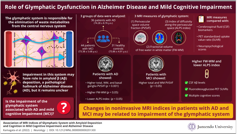
In a new study published in Neurology, a group of researchers including Christina Andica, Kaito Takabayashi, and Shigeki Aoki from Juntendo University in Japan and headed by Associate Professor Koji Kamagata from the Juntendo University Graduate School of Medicine explored the glymphatic system functioning in 36 patients with AD, 44 patients with mild cognitive impairment (MCI), and 31 healthy controls. The data was obtained from the Alzheimer’s Disease Neuroimaging Initiative-2 database.
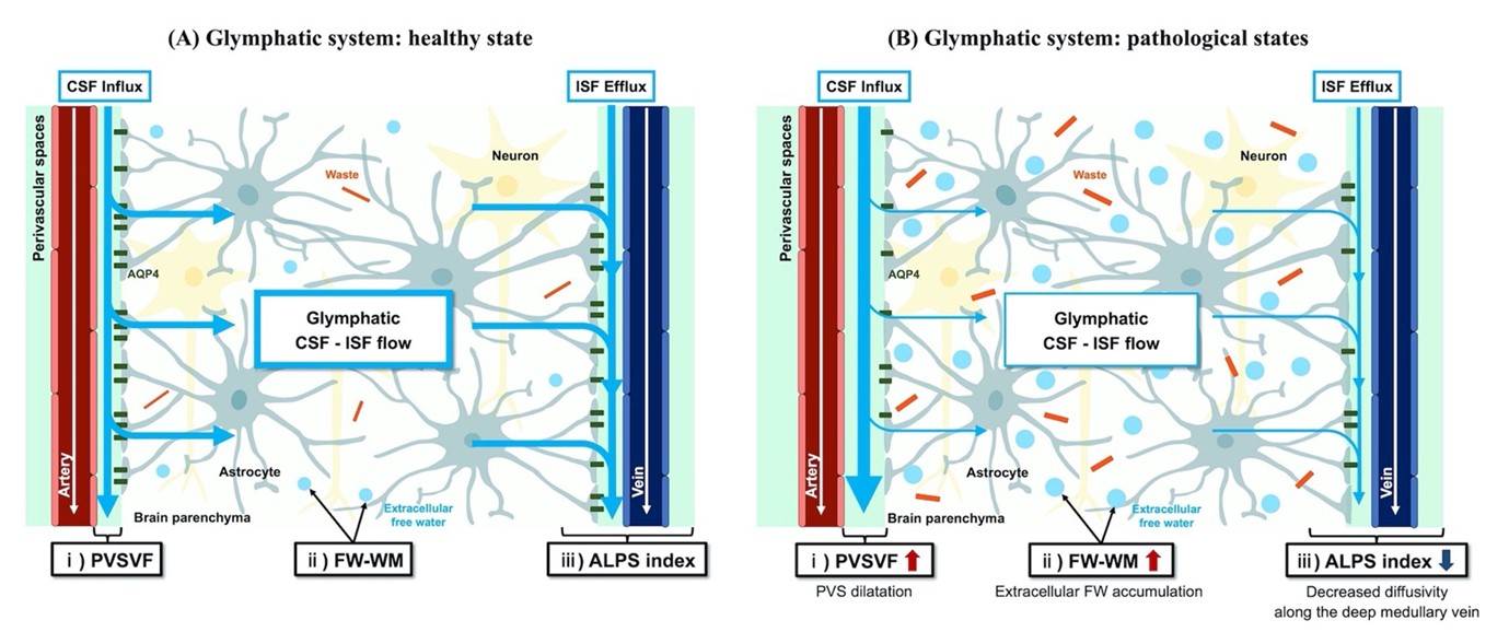
The group evaluated three MRI parameters of the glymphatic system in these patients— the volume of the perivascular space (PVS) in the brain, the fractional volume of free water in white matter (FW-WM), and the index of diffusivity along the perivascular space (ALPS index). “These three noninvasive indicators were chosen to detect malfunctions in the glymphatic system and correlate them with the levels of Aβ accumulation, neuropsychological score, and PET standardized uptake value ratio (SUVR),” says Dr. Kamagata. Their study was made available online on August 12, 2022 and published in the journal on September 19, 2022.
The results showed elevated PVSVF and volume of FW-WM, and a reduced ALPS index for patients with AD compared to the healthy controls, pointing towards a malfunction of the glymphatic system. Changes in the MCI patient group were only observed in PVSVF, suggesting early glymphatic impairment involving periarterial space. “In addition, our study also suggested associations between glymphatic dysfunction and Aβ deposition, neuronal damage, and cognitive impairment in AD,” highlights Dr. Kamagata.
These findings suggest that the impairment of the glymphatic system could indeed be associated with AD pathology and noninvasive MRI markers for monitoring its functioning could enable an early detection and treatment of AD.
The glymphatic system is the waste clearing pathway of the human brain. It is speculated that an abnormality in this system may be linked with the accumulation of amyloid beta (Aβ) proteins in our brain, a known pathological hallmark of Alzheimer disease (AD). A new study by researchers from Japan now show how an impaired glymphatic system in patients with AD may influence specific MRI indicators that can help track AD advancement in a noninvasive manner.
Basic concepts of the proposed MRI measures for indirect noninvasive evaluation of glymphatic system compartments in healthy and pathological states.
A new study by researchers from Japan has identified noninvasive MRI measurements for the glymphatic system that can be used as indicators to diagnose, prevent, and treat Alzheimer disease. Image credit: Dr. Koji Kamagata from Juntendo University, Japan.
License type: CC BY-NC-ND 4.0In a new study published in Neurology, a group of researchers including Christina Andica, Kaito Takabayashi, and Shigeki Aoki from Juntendo University in Japan and headed by Associate Professor Koji Kamagata from the Juntendo University Graduate School of Medicine explored the glymphatic system functioning in 36 patients with AD, 44 patients with mild cognitive impairment (MCI), and 31 healthy controls. The data was obtained from the Alzheimer’s Disease Neuroimaging Initiative-2 database.
The group evaluated three MRI parameters of the glymphatic system in these patients— the volume of the perivascular space (PVS) in the brain, the fractional volume of free water in white matter (FW-WM), and the index of diffusivity along the perivascular space (ALPS index). “These three noninvasive indicators were chosen to detect malfunctions in the glymphatic system and correlate them with the levels of Aβ accumulation, neuropsychological score, and PET standardized uptake value ratio (SUVR),” says Dr. Kamagata. Their study was made available online on August 12, 2022 and published in the journal on September 19, 2022.
The results showed elevated PVSVF and volume of FW-WM, and a reduced ALPS index for patients with AD compared to the healthy controls, pointing towards a malfunction of the glymphatic system. Changes in the MCI patient group were only observed in PVSVF, suggesting early glymphatic impairment involving periarterial space. “In addition, our study also suggested associations between glymphatic dysfunction and Aβ deposition, neuronal damage, and cognitive impairment in AD,” highlights Dr. Kamagata.
These findings suggest that the impairment of the glymphatic system could indeed be associated with AD pathology and noninvasive MRI markers for monitoring its functioning could enable an early detection and treatment of AD.
Basic concepts of the proposed MRI measures for indirect noninvasive evaluation of glymphatic system compartments in healthy and pathological states.
A new study by researchers from Japan has identified noninvasive MRI measurements for the glymphatic system that can be used as indicators to diagnose, prevent, and treat Alzheimer disease.
Image credit: Juntendo University, Japan.
License type: CC BY-NC-ND 4.0
Reference
| Authors | Koji Kamagata 1, Christina Andica 1,2, Kaito Takabayashi 1, Yuya Saito 1, Toshiaki Taoka 3, Hayato Nozaki 1, Junko Kikuta 1, Shohei Fujita 1, Akifumi Hagiwara 1, Kouhei Kamiya 3, Akihiko Wada 1, Toshiaki Akashi 1, Katsuhiro Sano 1, Mitsuo Nishizawa 1, Masaaki Hori 4, Shinji Naganawa 5, Shigeki Aoki 1 on behalf of the Alzheimer’s Disease Neuroimaging Initiative |
| Title of original paper | Association of MRI Indices of Glymphatic System With Amyloid Deposition and Cognition in Mild Cognitive Impairment and Alzheimer Disease |
| Journal | Neurology |
| DOI | 10.1212/WNL.0000000000201300 |
| Affiliations | 1. Department of Radiology, Juntendo University Graduate School of Medicine, Tokyo, Japan
2. Faculty of Health Data Science, Juntendo University, Chiba, Japan 3. Department of Innovative Biomedical Visualization (iBMV), Nagoya University Graduate School of Medicine, Nagoya, Japan 4. Department of Radiology, Toho University Omori Medical Center, Tokyo, Japan 5. Department of Radiology, Nagoya University Graduate School of Medicine, Nagoya, Japan |